For rehabilitation treatment plans caused by movement disorders due to neurological diseases, it is necessary to conduct preliminary animal experiments. These results are used to analyze the mechanisms of the disease and provide experimental evidence for devising treatment/rehabilitation plans.
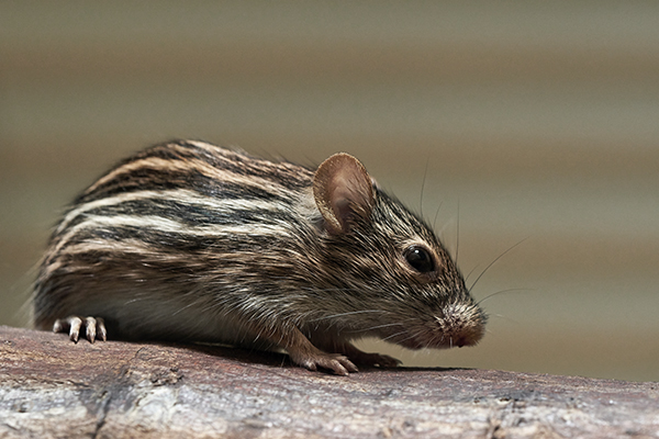
Spinal cord injury is a type of central nervous system damage that can lead to partial or complete loss of motor function. A primary focus of research on spinal cord injury is the recovery of residual motor function output, which can directly reflect the extent of injury or the effectiveness of treatment. One issue in this research is that some studies rely on subjective evaluations when assessing the recovery of motor function output, which can be insensitive and imprecise regarding changes in motor output.
Research Case Introduction
Gait analysis of spinal cord injury in rhesus monkeys - Beihang University
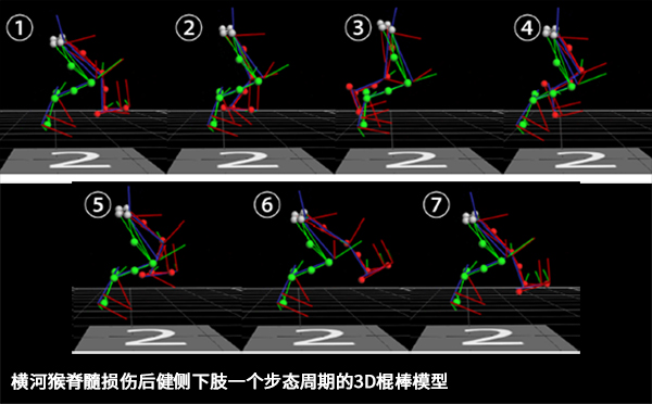
The research team from Beihang University chose an optical motion capture system to assist in their experiments. They conducted gait tests on rhesus monkeys before spinal cord injury, as well as 6 and 12 weeks after injury, in order to obtain more accurate and objective gait characteristics.
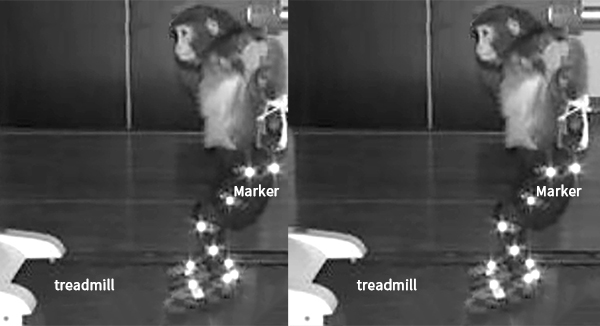
Before gait testing, a rhesus monkey is restrained in the same state as during the training period, with 16 reflective markers affixed to the lower limb joints (as shown in the figure above). Eight motion capture cameras are used to collect lower limb gait data at a frequency of 100Hz. During the test, the animal walks on a treadmill at 0.8 km/h, and data from continuous steps greater than 10 are retained. The collected data includes the duration of the double lower limb gait cycle, stride length, step height, the amplitude of knee/ankle joint angles, and the ratio of linkage parameters.
Parkinson's Disease Rat - Cardiff University
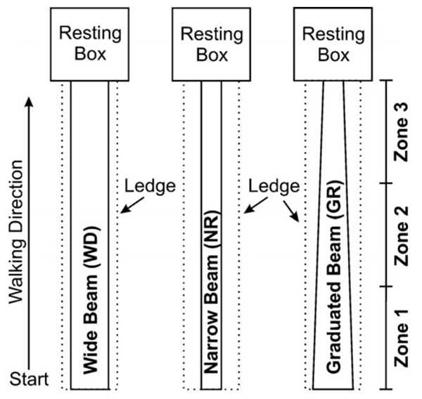
Similarly, researchers at Cardiff University, while studying the locomotion of rats with Parkinson's disease on beams of varying widths, chose to use optical motion capture equipment for movement analysis (as shown below).
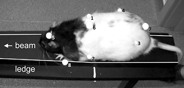
Results show that motion capture systems can provide quantified temporal gait parameters for rat models of Parkinson's disease, making it an effective method to study the impact of Parkinson's disease on movement. Researchers suggest that 3D motion capture technology can be used to collect data from animal disease models and compare it with human motion analysis. This helps to correlate human diseases with their respective animal models and explore the relationship between animal models and human behavior.
Mouse whisker motion patterns - University of Tennessee
Rodents such as rats and mice have sensitive tactile whiskers which they actively control to detect terrain. However, studies on the movement patterns of mouse whiskers are still in the early stages. Tracking the motion of whiskers is challenging due to their fast movement and fine structure.

The research team from the University of Tennessee utilized an optical motion capture system to attach tiny reflective markers to the whiskers of mice and fix the head with a specialized support, while a piece of cardboard was attached to the forepaws of the mice to prevent them from trying to remove the markers. This method of measuring mouse whisker movement is very precise and can be used for neurological and genetic research.
Monkey social brain psychology experiment - RIKEN Brain Science Institute
In addition to physiological experiments, researchers also conduct related psychological experiments by analyzing the behavior of intelligent primates. The social brain function allows our behavior to adapt to the social environment, but due to technical limitations, we know very little about how neurons recognize and regulate responses to social behavior. To address this field, researchers from the RIKEN Brain Science Institute designed experiments using two monkeys (M1 and M2) for their studies.
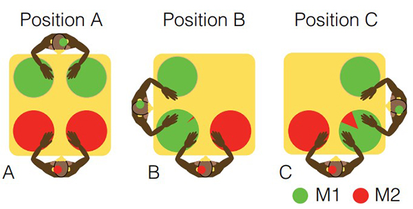
The experiment is divided into three scenarios, namely, two monkeys in non-overlapping spaces (A) and overlapping spaces (B and C). Food is placed at various locations on the table, and the monkeys' actions are observed under different circumstances. The lower body of the monkeys is fixed to the chairs, while their upper bodies can move freely. In the diagram above, the pie chart on the table represents the success rate of each corresponding colored monkey obtaining food when it is placed in that area. Tungsten electrodes have been implanted in the brains of the two monkeys to record neural activity, and they are dressed in specially made motion capture suits (with reflective markers attached to both shoulders, elbows, wrists, and hands, as well as the forehead and the back of the head), using an optical motion capture system to record the movements of the arms and head. The experiment only records arm movements with a speed higher than 30mm/s, as setting a threshold can better eliminate random movements. The motion capture system can help researchers achieve accurate quantitative analysis.
Whether the ultimate goal is to capture animal movement characteristics, analyze behavior, or simply record animal motion, NOKOV optical motion capture systems can provide suitable solutions. Below are some application cases from our customers.
Examples of NOKOV Motion Capture Projects
Sun Yat-sen University School of Medicine
In spinal cord rehabilitation assessment, recovery of the motor nervous system is an extremely important indicator. In experiments at the Sun Yat-sen University Medical School, the activity of the canine hind limb knee joint is primarily used as an external manifestation of the recovery of the motor nervous system.
In the experimental process, an area of approximately 3m*3m was constructed for dog activities, with eight NOKOV optical 3D motion capture cameras distributed around the area. Reflective markers were placed on the root of the dog's lower limbs, knee joints, and ankle joints, and the motion capture cameras were used to collect position information of these markers, thereby collecting data on the dog's joint movements within this space. By comparing the motion data of paralyzed dogs with that of normal dogs, it is possible to assess the recovery of the dog's motor nervous system and, consequently, the recovery of the dog's spinal cord.
References
[1] Zhao Can, Wei Ruihan, Zhao Wen, Ji Run, Rao Jiasheng, Yang Zhaoyang, Li Xiaoguang. Evaluation of Residual Stepping Ability in Spinal Cord Injured Rhesus Monkeys [J]. China Rehabilitation Theory and Practice, 2020, 26(06): 648-653.
[2] MadeteJK,KleinA,FullerA,etal.Challengesfacingquantificationofratlocomotionalongbeamsofvaryingwidths[J].ProceedingsoftheInstitutionofMechanicalEngineersPartHJournalofEngineeringinMedicine,2010,224(11):1257-65.
[3] SnigdhaR,BryantJL,CaoY,etal.High-Precision,Three-DimensionalTrackingofMouseWhiskerMovementswithOpticalMotionCaptureTechnology[J].FrontiersinBehavioralNeuroscience,2011,5(23):27.
[4] FujiiN,HiharaS,IrikiA.Dynamicsocialadaptationofmotion-relatedneuronsinprimateparietalcortex[J].PloSone,2007,2(4):e397.