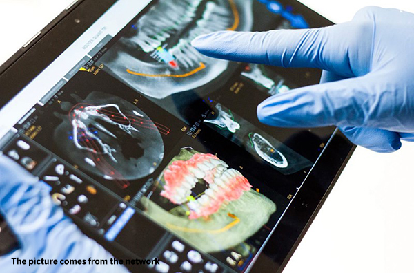
Since the first commercial CT came out in 1970s, CT technology has developed rapidly because of its great contribution to human health and great economic benefits in the field of industrial nondestructive testing. Nowadays, CT occupies an indispensable position in the field of clinical examination departments in hospitals, scientific research departments in universities and nondestructive testing in factories. Cone-beam CT has become a research hotspot in the field of CT due to the strong development of flat panel detectors.
Cone-beam CT system, called CBCT for short, is a typical three-dimensional imaging system. By emitting low-energy cone-beam rays, the rays and sensors can rotate around the patient or test object synchronously for one or less weeks, and the scanning process usually takes only ten seconds to tens of seconds. Cone beam CT has many advantages, such as high spatial resolution, high ray utilization rate, fast reconstruction speed, simple system and so on. It can be reconstructed by a variety of algorithms, among which FDK algorithm is widely used in practice because of its simple mathematical form, high computational efficiency and good reconstruction effect under the condition of small cone angle.

However, the FDK reconstruction algorithm has extremely high requirements for the system, which needs to meet three strict geometric alignment relationships:
1) the focus, rotation center and detector center of the radiation source are in a straight line;
2) The connecting line of the above three points is perpendicular to the plane of the detector;
3) The projection of the rotating shaft on the detector should coincide with the central column of the detector.
Therefore, accurate positioning of geometric parameters of cone-beam CT system is particularly important to improve the quality of reconstructed images.
In view of this key problem, Professor Niu Tianye's team from the School of Translational Medicine, Zhejiang University has effectively carried out animal experiments by using the cone beam CT system of small animals based on rotating gantry.

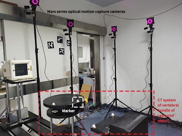
In this experiment, it is very important to correct the geometric position of cone-beam CT platform. Cone-beam CT system is mainly composed of ray source, workpiece turntable and area array detector, and the system geometric parameter error mainly comes from the installation deviation of these three parts. Because of the requirement of high precision for positioning correction, they chose NOKOV 3D optical motion capture system as the positioning tool.
The experimental team assigned the spatial coordinate origin and coordinate system to a specific position and direction on the CT platform, and arranged Markers on the X-ray source, workpiece turntable and area array detector of the CT platform to obtain coordinate data in real time, and corrected the cone beam CT platform by proofreading the data of the measured position.